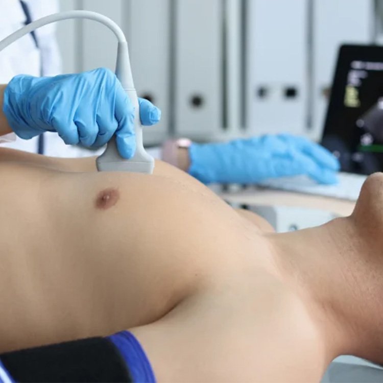
2D Echocardiogram
What is a 2D Echo (2D Echocardiogram)?
A 2D Echo (2D Echocardiogram) is a non-invasive ultrasound test that creates real-time images of the heart. This test uses high-frequency sound waves to produce a two-dimensional image of the heart, allowing doctors to examine the structure, function, and movement of the heart in detail. It is commonly used to assess the heart’s chambers, valves, walls, and blood flow.
How Does a 2D Echo Work?
During the procedure, a handheld device called a transducer is moved over your chest. The transducer emits sound waves that bounce off the heart’s structures and return to the machine, which processes the waves into live images. The procedure is painless and takes about 15 to 30 minutes.

Why is a 2D Echo Done?
A 2D Echo provides valuable information to help diagnose or monitor various heart conditions. It is commonly used to:
– Evaluate heart function and blood flow
– Detect abnormalities in the heart’s structure (such as enlarged heart chambers or thickened walls)
– Assess the condition of the heart valves and detect valve diseases
– Diagnose heart failure or cardiomyopathy
– Monitor the heart after a heart attack
– Check for congenital heart defects (especially in children)
What Do the 2D Echo Results Show?
The 2D Echo provides a detailed visual representation of your heart’s functioning. The test can reveal:
– Chamber size and thickness: Helps assess if the heart is enlarged or thickened.
– Heart valve function: Shows how well the valves open and close and whether there is any leakage (regurgitation) or narrowing (stenosis).
– Heart muscle motion: Checks how well the heart’s muscle contracts and relaxes.
– Blood flow: Assesses the speed and direction of blood flow within the heart.
Types of 2D Echo
There are several types of echocardiograms based on the area being examined and the specific condition:
– Transthoracic Echocardiogram (TTE): The standard 2D echo where the transducer is placed on the chest.
– Transesophageal Echocardiogram (TEE): A more detailed type where the transducer is passed down the esophagus to get clearer images of the heart, often used when more detail is needed.
– Stress Echocardiogram: A 2D echo performed while the heart is under physical stress (exercise or medication) to evaluate how the heart functions during activity.
Is a 2D Echo Safe?
A 2D Echo is a safe, non-invasive procedure that does not involve radiation. There are no known risks associated with the test, and it can be performed on individuals of all ages, including infants and pregnant women.
When Should You Get a 2D Echo?
Your doctor may recommend a 2D Echo if you experience symptoms such as chest pain, shortness of breath, swelling of the legs, or fatigue. It is also commonly performed as part of a routine evaluation for individuals with heart disease risk factors, such as high blood pressure, diabetes, or a family history of heart problems.
A 2D Echo is a critical tool in diagnosing heart conditions, enabling early detection and treatment to maintain optimal heart health.
